Thursday, February 21, 2019

Publication: Multi-directional optical coherence elastography

We recently published our first optical coherence elastography (OCE). In this research, we developed a new OCT based elastographic method, which visualizes microscopic tissue alteration induced by tiny (1-micron) compression of the tissue.

Among several variations of OCE, our method is characterized by its ability to determined the distribution of 3-D vectorial displacement and visualize local deformation of microscopic structure that is smaller than the optical resolution.

The method was applied to ex vivo porcine tissues, and successfully visualized microscopic mechanical response of the tissue.

The details are published in Optics Letters.

>> Full length article (Open access).

E. Li, S. Makita, S. Azuma, A. Miyazawa, and Y. Yasuno, "Compression optical coherence elastography with two-dimensional displacement measurement and local deformation visualization," Optics Letters 44, 787-790 (2019).

私達の研究グループにとって最初の光コヒーレンスエラストグラフィー(OCE)の論文を出版しました。この論文で報告した技術では、1ミクロンだけ計測組織を圧縮しながらトモグラフィー計測を行います。そして、そのトモグラフィー画像を解析することによって、この微小圧縮によって引き起こされる組織内部の変形の様子を可視化しています。

様々なバリエーションのあるOCE技術ですが、私達のOCTは組織の微小領域が三次元的にどの方向に移動しているのかを計測できることと、圧縮によって組織の中の光学分解能以下の組織の微小構造が変化している様子を可視化できることが特徴です。

今回の論文では、この手法を摘出された豚組織に適用し、圧縮による組織の機械的な応答を可視化することに成功しています。

この研究の詳細は米光学会のジャーナル Optics Letters に掲載されました。

>> Full length article (Open access).

E. Li, S. Makita, S. Azuma, A. Miyazawa, and Y. Yasuno, "Compression optical coherence elastography with two-dimensional displacement measurement and local deformation visualization," Optics Letters 44, 787-790 (2019).

Saturday, September 29, 2018

Yasuno gives BiOS Hot Topic Talk at SPIE Photonics West

私達の研究グループの安野が来年1月にサンフランシスコで開かれる国際会議 Photonics West / BiOS のプレナリーセッション(全員セッション)である BiOS Hot Topics において「マルチコントラスト OCT」を紹介します。

Photonics West / BiOS は 米光工学回(SPIE)が主催する世界最大の生体医用光学に関する学会です。BiOS Hot Topics セッションでは、生体医用光学の研究の最もホットなトピック8つを選び、それぞれの中心となる研究者がそれぞれの全体像を紹介するものです。

今回安野は、ホットな研究トピックとして、マルチコントラスト OCT/ Jones matrix OCT を紹介します。

詳細は SPIE/Photonics West のウェブサイトに掲載されています。

It is my honor to announce that Yasuno is going to give a plenary presentation at BiOS Hot Topic, SPIE Photonics West (San Francisco, January, 2019).

SPIE Photonics West / BiOS is the world largest conference conference of laser and biomedical optics. BiOS Hot Topic session is a plenary session, in which eight selected speakers introduce eight hottest topics in the year. Yasuno was designated to speak about recent activities on multi-functional optical coherence tomography.

The details of the session is available on  web-site of SPIE/Photonics West.


Publication: Pigment and flow imaging for posterior eye

Our colleague Shuichi Makita recently reported a new type of retinal optical coherence tomography (OCT), which visualizes the abnormality of retinal pigment epithelium (RPE) and vasculature in the eye. This OCT is referred to as ``pigment and flow imaging OCT'', or ``PAF-OCT'' in short. It uses a polarization diversity detection scheme to measure the polarization uniformity within the retina, and the polarization uniformity corresponds to the melanin distribution. In addition, the retinal flow is visualized by OCT angiography technique, simultaneously.

Since all optical fiber components are packed in a compact retinal scanner, it is good to use in an ophthalmic clinic. The PAF-OCT is, actually, now utilized in clinical investigation at Tokyo Medical University Ibaraki Medical Center. This compactness, easy operation and non-invasive ability to visualize the RPE and vascular abnormality make PAF-OCT a promising too for retinal diagnosis.

The details are reported in Biomedical Optics Express.

>>  Full length article (Open access)

S. Makita, T. Mino, T. Yamaguchi, M. Miura, S. Azuma, and Y. Yasuno, "Clinical prototype of pigment and flow imaging optical coherence tomography for posterior eye investigation," Biomed. Opt. Express 9, 4372–4389 (2018).

私達が新たに開発した網膜の色素・血流の三次元イメージング装置である「Pigment-and-flow Imaging Optical Coherence Tomography」 (PAF-OCT) について、米光学会の論文誌 Biomedical Optics Express に論文発表を行いました。この技術は、偏波分離検出メカニズムとOCTA(光断層干渉計アンギオグラフィー)技術を用いることにより、網膜内のメラニン色素の分布と血流の分布を三次元的に可視化するものです。これによって、外部からの外部からの薬剤の注入などを行わず、完全に非侵襲に網膜の色素上皮と循環系の異常を可視化することができます。

また、今回は発表した PAF-OCT 装置では、すべての機器コンポーネントをコンパクトな筐体(網膜スキャナー)の中に搭載することに成功しました。これにより、この装置は眼科医院などの臨床現場での安定稼働が可能になりました。実際に、現在この PAF-OCT 装置は東京医科大学茨城医療センター眼科で臨床試験を行っています。

PAF-OCT のもつこの安定性、容易な操作性、そして色素上皮と循環系の非侵襲可視化という機能は、加齢黄斑変性などの眼科疾患の検出に力を発揮すると考えられます。

この研究の詳細はアメリカ光学会の論文誌 Biomedical Optics Express に掲載されました。

>>  Full length article (Open access)

S. Makita, T. Mino, T. Yamaguchi, M. Miura, S. Azuma, and Y. Yasuno, "Clinical prototype of pigment and flow imaging optical coherence tomography for posterior eye investigation," Biomed. Opt. Express 9, 4372–4389 (2018).

Monday, July 2, 2018

Publication: Machine-learning based multi-contrast segmentation of optic nerve head

Our colleague Deepa Kasaragod recently published her new development on automatic tissue segmentation of human optic nerve head.

In this new method, she combined unsupervised and supervised machine learning technology to process multi-contrast three-dimensional tomography of human optic nerve head. The unsupervised method automatically groups the pixels of multi-contrast tomography based on its optical properties. And this pixel groups are used to train the subsequent supervised tissue classifier algorithm. The train classifier finally successfully highlights (segments) the beam-structure in the lamina cribrosa. So, finally, this fine segmentation was performed without manual supervision.

The details of this research is reported in Biomedical Optics Express.

>> Full length article (open access).
D. Kasaragod, S. Makita, Y.-J. Hong, and Y. Yasuno, ``Machine-learning based segmentation of the optic nerve head using multi-contrast Jones matrix optical coherence tomography with semi-automatic training dataset generation,'' Biomed. Opt. Express 9, 3220-3243 (2018).

私達の研究グループの Deepa Kasaragod 研究員が視神経乳頭のOCT画像の微小な組織を自動抽出する手法を開発しました。この手法は、Jones matrix OCT と呼ばれるそうちで得られる、組織の様々な光学特性を表す複数のトモグラフィーの情報と機械学習手法を組み合わせることで特定の組織部位を自動抽出するものです。今回の手法では、まず、「教師なし学習法」と呼ばれるアルゴリズムでトモグラフィー内のピクセルそれぞれを、それぞれ異なった組織によりわけます。そして、そのよりわけた結果を参考データとして、「教師なし学習」とよばれるアルゴリズムで「組織弁別器」と呼ばれるプログラムを自動最適化します。最終的に、この最適化された組織弁別器を用いて、視神経乳頭のなかにある篩状板、特にその中のビームと呼ばれる微小構造を自動抽出することに成功しました。

この技術の詳細は、アメリカ光学会発行の Biomedical Optics Express 誌に掲載されました。

>> Full length article (open access).
D. Kasaragod, S. Makita, Y.-J. Hong, and Y. Yasuno, ``Machine-learning based segmentation of the optic nerve head using multi-contrast Jones matrix optical coherence tomography with semi-automatic training dataset generation,'' Biomed. Opt. Express 9, 3220-3243 (2018).

Monday, June 25, 2018

Publication: Multi-contrast segmentation of pathological posterior eye

Our colleague Shinnosuke Azuma recently reported a brand-new method to automatically highlight retinal pigment epithelium (RPE) and choroid. This highlighting, i.e., segmentation multiple contrast information obtained by Jones matrix OCT, and does not use any morphological information. And hence, it is applicable not only to normal eyes but also severely damaged retina.

Based on this segmentation, he also established several en face visualization methods of eye pathology. Among several of them, melano layer thickness maps was found to have strong similarity of near-infrared fundus auto fluorescence (FAF) image. Owing to this similarity and depth resolution nature of OCT, we now can interpret the en face FAF image in three-dimensional manner.

The details of the methods and its clinical results are reported in Biomedical Optics Express.

>> Full length article (open access)
S. Azuma, S. Makita, A. Miyazawa, Y. Ikuno, M. Miura, and Y. Yasuno, ``Pixel-wise segmentation of severely pathologic retinal pigment epithelium and choroidal stroma using multi-contrast Jones matrix optical coherence tomography,'' Biomed. Opt. Express 9, 2955-2973 (2018).

当研究室の博士課程後期で勉強している東神之介さんが、Jones matrix OCT でえられる複数のコントラストを組み合わせることで眼底の特定の組織を検出・表示する手法を開発しました。この手法では、Jones matrix OCTで得られる様々なタイプの光特性を四則演算や閾値によって処理することで、色素上皮や脈絡膜実質部位の抽出を行っています。この手法は組織の形態に関する情報を用いいずに組織抽出を行っています。そのため、従来の組織抽出しようとはことなり、大きく形状が変化したような重篤な疾患眼でも組織の抽出を行うことができます。

また、抽出された色素上皮の形態から en face メラニン量マップも作成しました。このマップを近赤外眼底自発蛍光を比べると、この両者が極めて類似していることがわかりました。この類似性と、Jones matrix OCTの三次元計測特性を用いることで、近赤外眼底自発蛍光発生の様子を三次元的に理解することが可能になってきています。

この研究の詳細はアメリカ
光学会の論文誌 Biomedical Optics Express に掲載されています。

>> Full length article (open access)
S. Azuma, S. Makita, A. Miyazawa, Y. Ikuno, M. Miura, and Y. Yasuno, ``Pixel-wise segmentation of severely pathologic retinal pigment epithelium and choroidal stroma using multi-contrast Jones matrix optical coherence tomography,'' Biomed. Opt. Express 9, 2955-2973 (2018).

Monday, May 14, 2018

Publication: Polarization sensitive imaging of glaucoma surgery

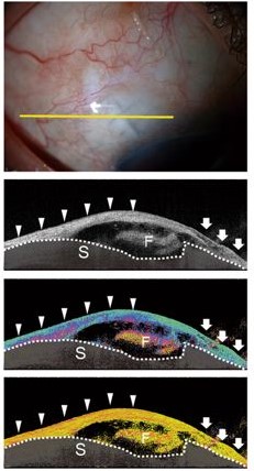
Our collaborator Shinichi Fukuda recently reported his comparative study among optical coherence tomography (OCT), conventional polarization sensitive OCT (PS-OCT's phase retardation imaging) and our new PS-OCT imaging method, i.e., birefringence imaging findings of bleb structure created in glaucoma surgery. As he concluded, the phase retardation imaging is particularly sensitive to slight alteration of bleb tissue, where as the birefringence imaging can highlight localized alteration of the tissue. He also found that the birefringence imaging can visualize "plate-like birefringence change" in the bleb, and it is associated with poor outcome of the surgery.

The details of study is described in Scientific Reports.

S. Fukuda, A. Fujita, D. Kasaragod, S. Beheregaray, Y. Ueno, Y. Yasuno, and T. Oshika, ``Comparison of intensity, phase retardation, and local birefringence images for filtering blebs using polarization-sensitive optical coherence tomography,'' Scientific Reports 8, 7519 (2018).

筑波大学眼科の福田慎一先生が、私達との共同研究である「緑内障手術痕の光干渉断層計(OCT)、偏光位相差トモグラフィー、複屈折トモグラフィー観察の比較」に関して Scientific Reports 誌で報告しています。この研究によって、偏光位相差トモグラフィーは緑内障手術痕における組織のごく弱い変化を検出することが可能であり、一方、複屈折トモグラフィーは組織の局所的な変化をとらえられることがわかりました。また、複屈折トモグラフィーで手術痕を観察したさい見られることのある「プレート状複屈折構造」が、手術予後不良を示す指標になることがしめされました。

この研究の詳細は Scientific Reports 誌で報告されています。


S. Fukuda, A. Fujita, D. Kasaragod, S. Beheregaray, Y. Ueno, Y. Yasuno, and T. Oshika, ``Comparison of intensity, phase retardation, and local birefringence images for filtering blebs using polarization-sensitive optical coherence tomography,'' Scientific Reports 8, 7519 (2018).

Friday, April 13, 2018

Opening for Student Positions

We are recruiting graduate (masters and PhD) and exchange students with scientific enthusiasm.  The students are expected to work for optical coherence tomography and microscopy. And the students will be fully or partially monetarily-supported. See the flyer for details. And contact Yasuno (yasuno@optlab2.bk.tsukuba.ac.jp)!

Links
Flyer for student recruit [PDF].

Tuesday, February 27, 2018

Publication: Eye-motion-corrected OCT angiography

Yiwei Chen recently reported our new results about a motion-free and non-invasive angiography method for living human eye. This method is based on OCT angiography (OCT-A) with newly designed special scanning pattern. The usage of Lissajous scan pattern is the key to achieve motion-free OCT-A. However, standard Lissajous scan is not compatible with OCT-A detection. So, Yiwei modified it to scan the same position on a tissue multiple times and enabled blood flow detection by OCT-A.  He further developed motion estimation and correction algorithms, successfully removed image distortion due to eye movements.

The details are reported in Biomedical Optics Express.

>>  Full length article (Open access)

Yiwei Chen, Young-Joo Hong, Shuichi Makita, and Yoshiaki Yasuno, ``Eye-motion-corrected optical coherence tomography angiography using Lissajous scanning,'' Biomed. Opt. Express 9, 1111-1129 (2018).


Yiwei Chen  研究員が私たちの最新の研究である「モーションフリー眼底血管イメージング手法」に関する論文を発表しました。これは、リサージュと呼ばれる連続曲線パターンで網膜を走査することで眼の動きによる画像の歪みを補正しながら、OCT造影(OCTA)と呼ばれる手法で眼底の血流を可視化する技術です。
 以前の研究で私たちは、リサージュ走査を用いた画像の歪みのない三次元眼底形状の撮影に成功していました。しかし、このリサージュ走査は OCTA と組み合わせることのできない操作方法でした。今回 Yiwei はリサージュ曲線の一部を、スキャンの連続性を維持しながら繰返すことで、OCTAで利用可能にしました。この技術を使うことで、眼球運動による歪みのない眼底血管分布を可視化することが可能になりました。

この研究の詳細はアメリカ光学会の論文誌 Biomedical Optics Express に掲載されました。

>>  Full length article (Open access)

Yiwei Chen, Young-Joo Hong, Shuichi Makita, and Yoshiaki Yasuno, "Eye-motion-corrected optical coherence tomography angiography using Lissajous scanning," Biomed. Opt. Express 9, 1111-1129 (2018) 

Tuesday, December 19, 2017

2017年度 応用物理特論 資料

筑波大学の講義「応用物理特論」(2017年度)で私たちのグループの研究室紹介を行いました。筑波大の学生さんだけでなく、私たちの研究に興味のある方のために資料を公開します。さらに興味のある方は RESEARCH ページと合わせて御覧ください。

>> 資料 PDF (6 MB)


Wednesday, December 13, 2017

研究生活入門

COGの日
皆さんこんにちわ!COGの研究管理補佐、長坂です!皆さんは理系の研究室の一日がどんなものか知っていますか?これまで理系とは縁のない生活を送ってきた私、長坂が驚きの理系研究室の一日を紹介します!


            9:00
            ~      出勤 
         11:00
      ・
                  ・
            ・      
       14:00    Standup meeting
                             Creaningtime (月曜日のみ)      
       14:15          論文
                     ・
                     ・
                     ・
       17:00
         ~           退勤
19:00


COGのプロジェクトの進め方
COGでは、Scrumというソフトウェア開発における手法を用いて研究を進めています。以下3つのイベントを1スプリント(期間の単位)の中で行い、繰り返し実施していきます。※COGでは、平日7日間で1スプリント
      
  ・Standup meeting・・・毎日14時から5分程度、
              1人一言 進捗自己確認と共有

  ・Review              ・・・スプリント終了の翌日、
               研究での結果について議論

  ・Retrospectiv    ・・・1、2スプリントに一度、
研究生活や進め方について意見を出し合う。


COGの主な研究発表スケジュール


 【研究発表目標】
   学会・・・国際学会1件、その他1件 発表
 
   論文・・・研究員は?
        修士の学生は、在学中にジャーナル論文1編 投稿
        博士の学生は、在学中にジャーナル論文2,3編 投稿


    4月   日本眼科学会
    5月 ARVO(アメリカ研究眼科学会)
    6月
    7月   Bios投稿締切
     8月
    9月 応用物理学会10月 臨床眼科学会
11月    OPJ
12月 ARVO投稿締切
1月 Bios
2月 卒論・修論
3月 

Tuesday, December 12, 2017

東神之介君がOPJ優秀講演賞を受賞しました!

当研究室の東神之介君が、11月に開催されたOptics & Photonics Japan(OPJ 日本光学会年次講演会)で第3回OPJ優秀講演賞を受賞しました。

東君は昨年の卒業研究で眼底検査用 Jones 行列OCTを開発し、今年4月の修士課程進学後は眼底Jones 行列OCTで得られる情報を用いた医療画像処理アルゴリズムの研究をしています。今回の受賞はその中でも眼底黄斑部の病変を可視化する画像処理手法に対するものでした。

東君にとって初めての学会発表での受賞となります。現在、東君はこの成果を英文論文として取りまとめています。東君の今後のさらなる活躍に期待しています!

受賞対象演題: "Macular analysis by multi-contrast segmentation of retinal pigment epithelium and choroid by Jones-matrix OCT"
>> 学会アナウンスサイト

偏光OCTレビューがトップダウンロードに選出されました

当研究グループの安野がウィーン医科大学の Christoph Hitzenberger 先生、アムステルダム自由大学の Johannes de Boer 先生と共同で執筆した論文 "Polarization sensitive optical coherence tomography - a review" が Biomedical Optics Express の最近一年で最もダウンロードされたOCT論文トップ 20 に選ばれました。Biomedical Optics Express はアメリカ光学会(OSA)の出版する論文誌であり、私たちの研究する生体光学のトップジャーナルの一つです。

この論文は私たちの研究グループが長年研究を続けている偏光感受型OCTの技術と医療応用を総覧する論文であり、これを読むだけでこの分野のことは一通り理解できるように書かれています。

簡単な論文の紹介と論文ウェイブサイトへのリンクは以前の記事にまとめられています。

Friday, November 10, 2017

Update: 私たちの研究

私たちの行っている研究を解説したページ "Research"  に2つのセクションを加えました。

  • Step 2. Our vision and project
  • Step 3. Further reading
です。Step 2. では今まさに私達が取り組んでいる研究プロジェクトについて解説しています。また、Step 3. では、さらに私たちの研究分野について勉強したい方のために教科書、論文を紹介しています。

ぜひ一度のぞいてみてください。

安野嘉晃

Friday, November 3, 2017

筑波大学 学園祭「学生プレゼンバトル」に安野が参加します

 2017年11月4日、筑波大学の学園祭(雙峰祭)の二日目に開催される「学生プレゼンバトル」企画で当研究のグループ・リーダーの安野が特別プレゼンテーションを行います。

学生プレゼンバトルとは、学群生や院生が自らの専攻している学問または研究の魅力を、 一般の聴衆に対してわかりやすく伝えるスキルを競う企画です。今回安野には、「そもそも研究活動ってどういうことなの?」という内容で、企画を主催する学生の方から依頼がありました。(ありがとうございます。)


ご興味のある方、学園祭にいらっしゃる予定の方、その他気になる方は、ぜひ、お越しください。


日時 :2017年11月4日 
    14:30~17:30(安野は16:15から特別プレゼンテーションを行います)
場所 : 大学会館3階ホール
テーマ:「社会の中の研究」
詳細は学生プレゼンバトルの web-site を御覧ください。

関連URL
雙峰祭 https://www.sohosai.tsukuba.ac.jp/(2017年11月3日~5日)
学生プレゼンバトル2017 http://tgn.official.jp/gpb2017/

追記: プレゼンスライド
プレゼンで使用したスライド(PDF)を公開します。プレゼンを見られなかった方、プレゼンの最後にでた「宿題」について考えたい方。ぜひダウンロードしてみてください。
>> スライド (PDF)

Sunday, October 29, 2017

研究発表: Optics Photonics & Japan 2017

日本光学会の年次講演会 Optics & Photonics Japan 2017 (2017/10/30 (月)-2017/11/2(木)、東京茗荷谷)において当研究室の学生・研究スタッフが4件の研究発表を行います。私(安野)が行う講演のように完成した研究をまとめて紹介するのではなく、いま私たちの研究室でもっともホットな研究を紹介します。

Four of our colleagues present their recent research at Optics & Photonics Japan 2017 (30th Oct. (Mon) to 2nd Nov. (Thu), Tokyo), an annual meeting of Optical Society of Japan. It is not like my (Yasuno’s) own invited presentations, but it covers cutting edge research topics now we are enthusiastically working for. I hope you can join it.


発表演題一覧 / List of presentations

11月1日(水)13:20-14:45 A会場「セッション: バイオ光学・バイオフォトニクス、医用光学(2)」
  • [1pA3] En Li et al., “Birefringence and deformation estimation of biological tissue by polarization sensitive optical coherence elastography” (偏光感受型光コヒーレンスエラストグラフィーによる生体組織の複屈折と微小変形の同時計測)
  • [1pA4] Shinnosuke Azuma “Macular analysis by multi-contrast segmentation of retinal pigment epithelium and choroid by Jones-matrix OCT” (ジョーンズ行列トモグラフィーのマルチコントラスト情報を用いた色素上皮・脈絡膜のセグメンテーション)
  • [1pA5] Deepa Kasaragod “Jones matrix optical coherence tomography based multi-contrast tissue classification of the posterior eye” (ジョーンズ行列トモグラフィーによる眼底組織弁別)
  • [1pA6] Shuichi Makita “Laser coagulation control by detecting tissue alteration with optical coherence tomography” (光コヒーレンストモグラフィーによる光凝固術における組織編成のモニタリングと凝固のアクティブ制御)
UPDATE
2017/11/1 (Wed) 全件、無事に発表できました。聞いていただいた皆様、質疑にご参加いただいた皆様、ありがとうございました。(安野)