The details of study is described in Scientific Reports.
S. Fukuda, A. Fujita, D. Kasaragod, S. Beheregaray, Y. Ueno, Y. Yasuno, and T. Oshika, ``Comparison of intensity, phase retardation, and local birefringence images for filtering blebs using polarization-sensitive optical coherence tomography,'' Scientific Reports 8, 7519 (2018).
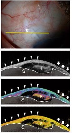
筑波大学眼科の福田慎一先生が、私達との共同研究である「緑内障手術痕の光干渉断層計(OCT)、偏光位相差トモグラフィー、複屈折トモグラフィー観察の比較」に関して Scientific Reports 誌で報告しています。この研究によって、偏光位相差トモグラフィーは緑内障手術痕における組織のごく弱い変化を検出することが可能であり、一方、複屈折トモグラフィーは組織の局所的な変化をとらえられることがわかりました。また、複屈折トモグラフィーで手術痕を観察したさい見られることのある「プレート状複屈折構造」が、手術予後不良を示す指標になることがしめされました。
この研究の詳細は Scientific Reports 誌で報告されています。
S. Fukuda, A. Fujita, D. Kasaragod, S. Beheregaray, Y. Ueno, Y. Yasuno, and T. Oshika, ``Comparison of intensity, phase retardation, and local birefringence images for filtering blebs using polarization-sensitive optical coherence tomography,'' Scientific Reports 8, 7519 (2018).