Wednesday, May 31, 2017

Publication: Review article for polarization sensitive optical coherence tomography

Our colleague Yasuno recently published an review article of "polarization sensitive optical coherence tomography (PS-OCT)," which is co-authored with Prof. Johannes de Boer (VU University of Amsterdam) and Prof. Christoph Hitzenberger (Medical University of Vienna).

This article provides a comprehensive overview of PS-OCT principle, hardware implementation, signal processing, and clinical applications. If you are interested in building or using PS-OCT, this article can be a good textbook. For beginners of OCT, this article can be a good introduction of
a functional extension of OCT.

>> Full length article (open access)

Citation: J. F. de Boer, C. K. Hitzenberger, and Y. Yasuno "Polarization sensitive optical coherence tomography – a review [Invited]," Biomed. Opt. Express 8, 1838-1873 (2017).

当研究グループの安野がアムステルダム自由大学の Prof. Johannes de Boer、ウィーン医科大学の Prof. Christoph Hitzenberger と共同で偏光感受型光コヒーレンストモグラフィー (偏光OCT)のレビュー論文を発表しました。

この論文は、偏光OCTの原理、ハードウェア実装、信号処理、臨床応用までを広く解説しています。偏光OCTの作成、利用に興味のある方には最初に読む教科書として最適なものになっていると思います。また、偏光OCTにまずは興味がある、という方にも 最初に読む文献として適切なものに仕上がっていると思います。

>> Full length article (open access)

Citation: J. F. de Boer, C. K. Hitzenberger, and Y. Yasuno "Polarization sensitive optical coherence tomography – a review [Invited]," Biomed. Opt. Express 8, 1838-1873 (2017).

Monday, May 1, 2017

Publication: Motion-free eye imaging by Lissajous OCT

Our colleague Yiwei Chen has reported a new scanning technology for retinal optical coherence tomography (OCT). He used a Lissajous scan pattern to scan in vivo human retina. The Lissajous scan has significant redundancy as it scans the eye multiple times but with low density for each time. This is a perfect property to compute and estimate the eye motion occurred during the scan. By using this estimated eye motion, we correct the image deformation of the OCT occurred by the eye motion.

This technology enables motion-free OCT imaging of the eye, by which we obtain a true morphological shape of the eye. Since myopia and glaucoma are associated with eye deformation, our Lissajous OCT might have a large impact on myopia and glaucoma research/diagnosis in the future.

The details are reported in Biomedical Optics Express. (See the bottom for the link).

私達の研究グループの Yiwei Chen 研究員がモーションフリー3次元眼底イメージングを実現するための技術である「リサージュOCT」を開発史、論文発表を行いました。この技術は、リサージュと呼ばれる連続曲線パターンでOCTのプローブ光をスキャンすることで眼底の構造情報を計測します。リサージュパターン粗い密度で何度も連続的に眼底をスキャンすることを特徴としています。この特性を活かすことで、撮影中に発生した眼球の動きを算出し、OCT画像に現れる動きの影響を除去することができます。この技術を使うことで、眼球の真の形状を計測することが可能になりました。

緑内障や近視などの疾患は、眼球の形状変化を伴うことが知られています。今回開発されたリサージュOCTを用いることにより、眼球の形状の変化を高精度に追跡することが可能になります。これにより、緑内障、近視の研究や診断手法の発展が期待されます。

この研究の詳細はアメリカ光学会の論文誌 Biomedical Optics Express に掲載されました。

>> Full-length article (open access)
C. Yiwei, Y.-J. Hong,  S. Makita, and Y. Yasuno, "Three-dimensional eye motion correction by Lissajous scan optical coherence tomography," Biomed. Opt. Express 8, 1783-1802 (2017).

Wednesday, March 1, 2017

Publication: Multi-contrast Jones matrix OCT for skin imaging

Our colleague En Li recently reported our
development on dermatological Jones matrix optical coherence tomography (JM-OCT). This is the first full-contrast OCT for skin imaging. It is based on 1.3-um swept-source JM-OCT principle and can 3-dimensionally and non-invasively visualize the structure, vasculature, collagen and melanin of human skin by a single shot.

The technical details and skin measurement examples are published in Biomedical Optics Express.

>> Full-length article (open access)
E. Li, S. Makita, Y.-J. Hong, D. Kasaragod, and Y. Yasuno, "Three-dimensional multi-contrast imaging of in vivo human skin by Jones matrix optical coherence tomography," Biomed. Opt. Express 8, 1290-1305 (2017).

博士課程で勉強している李恩  (LI, En) さんが皮膚計測用マルチコントラスト Jones matrix 光コヒーレンストモグラフィー (JM-OCT) のに関する論文を出版しました。この論文で報告されている JM-OCT は世界で最初の皮膚科学用途に特化した Jones matrix 型 OCTです。

この装置を使うことで、一度の計測で非侵襲に生きたヒト皮膚の構造、血管、コラーゲン、メラニンの3次元形態を可視化することができます。

装置の詳細、in vivo ヒト皮膚計測結果はアメリカ光学会の論文誌 Biomedical Optics Express に掲載されています。

>> Full-length article (open access)
E. Li, S. Makita, Y.-J. Hong, D. Kasaragod, and Y. Yasuno, "Three-dimensional multi-contrast imaging of in vivo human skin by Jones matrix optical coherence tomography," Biomed. Opt. Express 8, 1290-1305 (2017).

Wednesday, February 1, 2017

Publication: Quantitative birefringence estimator for Jones matrix OCT

Our colleague Deepa Kasaragod has reported her new mathematical theory and numerical algorithm that give highly quantitative measurement capability to Jones matrix optical coherence tomography (JM-OCT). And hence, it enabled three-dimensional quantitative measurement of in vivo tissue birefringence.

This method is based on a maximum a-posteriori estimation theory. It first compute the probability distributions of a true signal-to-noise ratio and a true birefringence when particular measurements of a birefringence and signal-strength were occurred. The estimator finally gives the most probable true birefringence value under the measured birefringence and signal-strength by using the probability distributions.

The method was validated numerically and also experimentally by investigating in vivo human eye.
The details are reported in Biomedical Optics Express (see the bottom for a link).

当研究室の Deepa Kasaragod 研究員が新しい複屈折推定理論・アルゴリズムに関する論文を発表しました。この理論・アルゴリズムは、Jones matrix OCT で得られた「計測された複屈折値」と「計測された散乱信号強度」から「もっとも確からしい真の複屈折値」を推定するものです。これによって生体複屈折の3次元定量計測が可能になりました。

この方法は「事後分布推定」と呼ばれる理論にもとづいています。あらかじめ、特定の複屈折と信号強度が計測された際に予想される「真の複屈折強度」の確率分布を計算しておきます。そして、実際の計測後に、その確率分布から計測された試料の真の複屈折を推定する、というものです。

この手法の理論、実装の詳細と数値的検証結果、生きたヒト眼底計測に適用した結果はアメリカ光学会の論文誌 Biomedical Optics Express に掲載されています。

>> Full-length article (open access)
D. Kasaragod, S. Makita, Y.-J. Hong, and Y. Yasuno, "Noise stochastic corrected maximum a posteriori estimator for birefringence imaging using polarization-sensitive optical coherence tomography," Biomed. Opt. Express 8, 653-669 (2017).


Tuesday, December 16, 2014

研究室紹介

COGでの研究活動に興味のある皆様へ。
Computational Optics Group の研究紹介スライドを公開します。

Tuesday, July 1, 2014

Publication: Accurate Quantification of Tissue Birefringence

Microstructures support the mechanical property of a tissue. And hence, investigating tissue microstructure has been a great interest both in the biomechanical research and for investigation of diseases associated with biomechanics, such as glaucoma. However, there was no way for in vivo quantification of tissue biomechanics. One promising approach, as we believe, is quantifying tissue birefringence for quantifying biomechanics. This approach is based on the fact that both the tissue biomechanics and birefringence are based on the same aspect of the tissue, i.e., microstructure. Our group has been working for polarization sensitive optical coherence tomography (PS-OCT) for the quantification of the biomechanics through quantitative measurement of tissue birefringence.

Since its innovation in 1997, PS-OCT was nice to qualitatively visualize the tissue birefringence. However, its quantification ability was low. It was mainly because its non-linear and complex effects of measurement noise into the birefringence values to be measured. To be honest, the raw birefringence value measured by PS-OCT has not been reliable at all.

Our colleagues Deepa Kasaragod recently developed a mathematical framework to accurately estimate the tissue birefringence from the raw birefringence values measured by PS-OCT. She first numerically characterized the relationship between the measurement noise and measured birefringence values. And then, she designed a mathematical frame work, by using a Bayesian rule together with the numerically obtained property, to obtain a “maximum likelihood estimation” of the tissue birefringence.

Both numerical and experimental validations proved that this framework has high ability to quantitate the birefringence. This method was recently published in Optics Express (full citation is shown below). In this paper, some in vivo human eye results including a pathologic case (anterior eye of trabeculectomy bleb) are presented.

Currently, a clinical study with this method is ongoing. I believe we can present impressive clinical results soon. The details of the technology are now available on the following paper.

Full length article (open access)
D. Kasaragod, S. Makita, S. Fukuda, S. Beheregaray, T. Oshika, and Y. Yasuno, Optics Express 22, 16472-16492 (2014), http://dx.doi.org/10.1364/OE.22.016472

Joschi, Yoshiaki Yasuno

Thursday, October 18, 2012

Visualization of Choriocapillaris by Doppler OCT

Our colleague Kazuhiro Kurokawa reported in vivo imaging of choriocapillaris by using Doppler optical coherence tomography (Doppler OCT) equipped with adaptive optics (AO) retinal scanner. Doppler OCT have been long time utilized for the investigation of retinal vasculature. Despite of its high ability for the visualization of vasculature, it was not possible to visualize choriocapillaris mainly because of its very small dimensions. We overcame this issue by using a custom made AO retinal scanner.
      The details are presented in our recent paper in Optics Express.

>> Full length article (open access)
Citation: K. Kurokawa, K. Sasaki, S. Makita, Y.-J. Hong, and Y. Yasuno, "Three-dimensional retinal and choroidal capillary imaging by power Doppler optical coherence angiography with adaptive optics," Opt. Express 20, 22796-22812 (2012).

Scleral biomechanics and birefringence

Our colleague Masahiro Yamanari recently reported a new method for noninvasive assessment of scleral biomechanics. The biomechanics of sclera is supported by its micro-structure that is mainly formed by collagen fibers. Since the collagen fibers is known to have strong form birefringence, we may be able to assess the biomechanics by measuring the birefringence. Yamanari demonstrated significant correlation between the scleral mechanical stiffness and its birefringence which was measured by a polarization sensitive optical coherence tomography.
      You can find more details in our article on PLoS ONE.

>> Full length article on journal web site (open access)
Citation: M. Yamanari, K. Ishii, S. Fukuda, Y.H. Lim, S. Makita, M. Miura, T. Oshika, and Y. Yasuno, "Optical rheology of porcine sclera by birefringence imaging," PLoS ONE 7, e44026 (2012).

Joschi, Yoshiaki Yasuno

Quantifying Blood Flow in Choroid

Figure Miura 2012 IOVS.Our colleague Masahiro Miura from Tokyo Medical University recently reported our first trial of quantification of choroidal blood flow. He utilized a custom made Doppler optical coherence tomography with a probe beam at 1.0-um band. The Doppler signal at the choroid was further processed with a structural structural information of the vessel. Finally an absolute velocities of choroidal blood flow in in vivo human eyes were presented.
     The details are presented in a recent issue of Investigative Ophthalmology and Visual Science.

>> Abstract and full length article on the journal web-site

Citation: M. Miura, S. Makita, T. Iwasaki, Y. Yasuno, "An approach to measure blood flow in single choroidal vessel using Doppler optical coherence tomography," Invest. Ophthalmol. Vis. Sci. 53, 7137-7141 (2012).

Wednesday, August 15, 2012

Step-by-step method for scientific paper writing

Yasuno recently released a short instruction article of a step-by-step method to write a scientific paper. This article was originally written to teach his students the way of scientific writing. In this instruction, he discusses a purpose of scientific writing and a detailed way to construct a logic of the paper.

The instruction is distributed under a Creative Commons Attribution No Derivatives 3.0 License (CC BY-ND 3.0), and hence you can freely read it, use it for your teaching, and also can distributed. Although you don't need to obtain any permission to use/re-distribute this document, any criticism and positive feedback are also welcome.

The document is available on the following distribution site.
>> A Card-Based Method for Scientific Paper Writing

2012-08-15
Joschi, Yoshiaki Yasuno

Monday, December 26, 2011

Journal paper: Choroidal optical coherence angiography with variable velocity range

Non-invasive and three-dimensional angiography of choroid with several blood-velocity range is available now. Our colleague Franck Jaillon recently published the details of this methodology in Optics Express. In this paper he demonstrated non-invasive visualization of fine vasculature of the choroid of normal eyes. This technology is based on Doppler optical coherence tomography, and hence it is totally noninvasive and three-dimensional. The Doppler detection is performed with our original dual beam scan Doppler scheme, which provides extremely high Doppler sensitivity. In addition, this system uses a 1-um probe beam, it provides deep penetration into the choroid.

   The details are described in the following paper.
>> Full length article on Journal web-site (open access)
Citation: F. Jaillon, S. Makita, Y. Yasuno, "Variable velocity range imaging of the choroid with dual-beam optical coherence angiography," Opt. Express 20, 385-396 (2012).

Tuesday, July 26, 2011

Journal Article: Office-Based Polarization Sensitive OCT for Anterior Eye

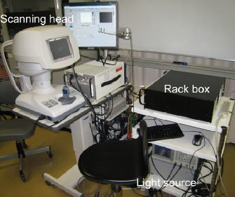
Our colleague Yiheng Lim recently reported a clinical prototype of polarization sensitive optical coherence tomography (PS-OCT) specially designed for the investigation of anterior eye segments. This system based on our developed high-speed Jones matrix OCT methodology, and integrated with a scanning head specially designed for anterior eye imaging. This clinical prototype provides semi-automatic alignment function and possesses a compact, office-compatible size. Several clinical cases were investigated to prove the clinical utility.

The details are reported in Biomedical Optics Express.

>> Full length article on journal web-site (open access)

Citation: Y.H. Lim, M. Yamanari, S. Fukuda, Y. Kaji, T. Kiuchi, M. Miura, T. Oshika, and Y. Yasuno "Birefringence measurement of cornea and anterior segment by office-based polarization-sensitive optical coherence tomography," Biomed. Opt. Express 2, 2392-2402 (2011).

Sunday, April 10, 2011

Journal Article: High-Sensitive Doppler Imaging of Choroid by Dual-Beam Doppler OCT

Our colleague Franck Jaillon recently reported his new Doppler optical coherence tomography (OCT) system which enables high-penetration and dual-velocity imaging of choroidal vasculature. This methodology employed   newly developed dual-beam OCT scanning head to provide high-sensitive Doppler imaging. Furthermore, this scanning head can provide multiple velocity range for Doppler detection. Since this system is using 1-um light as a probe, the details of in vivo choroidal vasculature was successfully visualized.

The details are published in Biomedical Optics Express.

>> Full length article on journal web-site (open access)

Citation: F. Jaillon, S. Makita, E.-J. Min, B.H. Lee, Y. Yasuno, "Enhanced imaging of choroidal vasculature by high-penetration and dual-velocity optical coherence tomography," Biomed. Opt. Express 2, 1147-1158 (2011).

Thursday, October 14, 2010

Journal Article: Wettability Characterization by OCT

Tapio Fabritius (Oulu University, Finland) recently published a paper in Optics Express in which the authors investigated the utility of optical coherence tomography (OCT) for the characterization of wettability of materials. The study presented was a collaboration between Oulu University and Computational Optics Group at the University of Tsukuba.
In the particular demonstration in this paper, a glycerol droplet on a rough surface was characterized, and the contact angles between them were automatically obtained.

The details are published in the following Optics Express paper.

Wednesday, June 16, 2010

Journal Article: Full Range Polarization Sensitive OCT at 1-Micron

Our colleague Masahiro Yamanari recently reported a new polarization sensitive optical coherence tomography (PS-OCT) for retinal and choroidal imaging. This new PS-OCT utilized a probe beam with 1-micron wavelength enabled high-penetration to the choroid, and a full range measurement scheme based on the modulation of reference beam of OCT (BM-scan). This system enables detailed investigation of birefringence property of the deep posterior eye.
After all, this OCT possesses a depth measurement range of 5.3 micrometers in air, a depth resolution of 11.4 micrometers in tissue, and a measurement speed of 30,000 depth-scan/sec. The intensity ratio between an OCT signal and its suppressed mirror signal was found to be 40.5 dB.
This PS-OCT is the first demonstration of 1-um PS-OCT with a clinically useful depth measurement range.
The details are available in the following Optics Express paper.

Citation: M. Yamanari, S. Makita, Y. Lim, and Y. Yasuno, "Full-range polarization-sensitive swept-source optical coherence tomography by simultaneous transversal and spectral modulation," Opt. Express 18, 13964-13980 (2010).