Tuesday, October 17, 2023

Publication: Glaucoma diagnosis can be affected by polarization artifacts of OCT

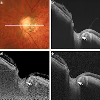
Our collaborator, Masahiro Miura, recently revealed significant artifacts exist in optical coherence tomography (OCT) images of the lamina cribrosa (LC).

The LC is the mechanically stiff tissue at the bottom of the eyeball, supporting the internal pressure of the eye. Abnormalities in the LC are known to be highly related to the risk of glaucoma. Given that glaucoma is one of the leading causes of blindness, the assessment of the LC is of significant importance. While it has been believed that defects in the LC can be visualized by OCT, Miura found that a significant proportion (5.9% of cases) of the LC defects found in OCT are not actual defects but rather artifacts caused by the unauthorized effect of light polarization.

This finding may impact glaucoma diagnosis, but we believe that such artifacts can be corrected by using our developing polarization-sensitive OCT.

The details of this research are published in Scientific Reports.


Citation:
M. Miura, S. Makita, Y. Yasuno, H. Nakagawa, S. Azuma, T. Mino, A. Miki, “Birefringence-derived artifact in optical coherence tomography imaging of the lamina cribrosa in eyes with glaucoma,” Sci. Rep. 13, 17189 (2023). https://doi.org/10.1038/s41598-023-43820-5

>> Full article (open access)

わたしたちの共同研究者である三浦雅博さんが網膜の篩状板の光コヒーレンストモグラフィー(OCT)像に著しいアーチファクトが存在することを明らかにしました。

篩状板は眼球の底にある硬い組織であり、眼球内部からの圧力を支えています。篩状板の異常は緑内障のリスクに大きく関係していることが知られており、また、緑内障が失明の主要な原因の一つであることを考えると、篩状板の評価は眼科診療において重要な意味を持ちます。これまで篩状板の異常(欠損)はOCTで可視化できていると考えられてきましたが、今回の研究により、OCTで発見された篩状板の欠陥のかなりの割合(調査した症例の5.9%)が、実際の欠損ではなく、偏光の影響によるアーチファクトであることがわかりました。

この知見は緑内障診断に影響を与える可能性がありますが、我々が開発している偏光感受型OCTを用いれば、このようなアーチファクトを補正できると考えています。

本研究の詳細はScientific Reports誌に掲載されています。

Citation: M. Miura, S. Makita, Y. Yasuno, H. Nakagawa, S. Azuma, T. Mino, A. Miki, “Birefringence-derived artifact in optical coherence tomography imaging of the lamina cribrosa in eyes with glaucoma,” Sci. Rep. 13, 17189 (2023). https://doi.org/10.1038/s41598-023-43820-5

>> Full article (open access)

(この記事の日本語版は www.DeepL.com で翻訳したものを一部修正したものです。)
Monday, October 16, 2023

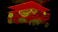
Publication: Dynamic OCT reveals spatial response of anti-cancer drug in spheroids

 Our colleague, Ibrahim Abd El-Sadek, recently revealed that the spatial pattern induced by anti-cancer drugs in tumor spheroids varies among tumor types and drug types.

Ibrahim applied his own developed dynamic optical coherence tomography (D-OCT) method to 3D tomographic visualization of tumor spheroids treated by anti-cancer drugs. The study involved two types of cancer cells, including breast cancer cells (MCF-7) and colon cancer cells (HT-29), and the spheroids were treated by two types of anti-cancer drugs, including paclitaxel (PTX, Taxol) and SN-38. The D-OCT revealed that the size, shape, and cellular activity are highly affected by anti-cancer drugs, and the spatial pattern of cellular activity alteration also depends on the cell type and the drug type. 

The details of this research are published in Scientific Reports.

Citation: I.A. El-Sadek, L.T.W. Shen, T. Mori, S. Makita, P. Mukherjee, A. Lichtenegger, S. Matsusaka, and Y Yasuno, “Label-free drug response evaluation of human derived tumor spheroids using three-dimensional dynamic optical coherence tomography,” Sci. Rep. 13, 15377 (2023). https://doi.org/10.1038/s41598-023-41846-3

>> Full article (open access)


わたしたちの研究室の Ibrahim Abd El-Sadek さんが腫瘍スフェロイドにおいて抗癌剤によって誘導される空間パターンが、腫瘍の種類や薬剤の種類によって異なることを明らかにしました。

El-Sadek さんは独自に開発した dynamic 光コヒーレンストモグラフィー法(D-OCT)を抗がん剤で処理した腫瘍スフェロイドの3D断層可視化に応用しました。この研究では、乳がん細胞(MCF-7)と結腸がん細胞(HT-29)の2種類のがん細胞のスフェロイドを用い、それらのスフェロイドをパクリタキセル(PTX、タクソール)とSN-38を含む2種類の抗がん剤で処理しました。D-OCT撮影の結果、スフェロイドの大きさ、形状、細胞活性が抗がん剤に大きく影響され、さらに、細胞活性の変化の空間パターンも細胞の種類や薬剤の種類によって異なることが明らかになりました。

この研究の詳細はScientific Reports誌に掲載されています。

Citation: I.A. El-Sadek, L.T.W. Shen, T. Mori, S. Makita, P. Mukherjee, A. Lichtenegger, S. Matsusaka, and Y Yasuno, “Label-free drug response evaluation of human derived tumor spheroids using three-dimensional dynamic optical coherence tomography,” Sci. Rep. 13, 15377 (2023). https://doi.org/10.1038/s41598-023-41846-3

>> Full article (open access)

(この記事の日本語版は www.DeepL.com で翻訳したものを一部修正したものです。)

Sunday, October 15, 2023

Publication: Novel label-free imaging technique reveals kidney function

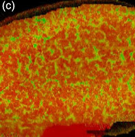
Our colleague Pradipta Mukherjee reported on a study that uncovers the intricated metabolic and functional structure of kidneys using our developed dynamic optical coherence tomography (DOCT) method.

The metabolic function of the kidney is pivotal to kidney health and susceptible to renal disease. This research introduces a three-dimensional label-free imaging technique, DOCT, to observe dynamic renal activity in normal and obstructed mouse kidney models ex vivo.

In the DOCT images of normal kidneys, the renal tubules appeared as convoluted pipe-like structures, while such structures were not observed in the obstructed kidneys.  This study suggests that DOCT can be a useful tool to non-invasively investigate renal structures and functions

The details of this research are published in Scientific Reports.

Citation: Pradipta Mukherjee, Shinichi Fukuda, Donny Lukmanto,  Thi Hang Tran, Kosuke Okada, Shuichi Makita, Ibrahim Abd El-Sadek, Yiheng Lim, and Yoshiaki Yasuno "Renal tubular function and morphology revealed in kidney without labeling using three-dimensional dynamic optical coherence tomography," Sci. Rep. 13, 15324 (2023). https://doi.org/10.1038/s41598-023-42559-3

わたしたちの研究室の Pradipta Mukherjee さんが わたしたちの開発している dynamic optical coherence tomography (DOCT) 法を用いて腎臓の代謝と機能の非侵襲な可視化に成功しました。
腎臓の代謝機能は、腎臓の健康にとって極めて重要であり、腎臓病により強く影響を受けます。本研究では、3次元ラベルフリーイメージング技術であるDOCTを用いることで、正常および閉塞モデルマウスの腎臓の活動の ex vivo 可視化を行いました。
正常な腎臓のDOCT画像では、腎尿細管が複雑なパイプ状構造として観察されました。一方、閉塞モデル腎ではそのような構造が消失することがわかりました。 この研究により、DOCTが腎臓の構造と機能を非侵襲的に調べるための有用なツールであることが示されました。
この研究の詳細はScientific Reports誌に掲載されています。
Citation: Pradipta Mukherjee, Shinichi Fukuda, Donny Lukmanto,  Thi Hang Tran, Kosuke Okada, Shuichi Makita, Ibrahim Abd El-Sadek, Yiheng Lim, and Yoshiaki Yasuno "Renal tubular function and morphology revealed in kidney without labeling using three-dimensional dynamic optical coherence tomography," Sci. Rep. 13, 15324 (2023). https://doi.org/10.1038/s41598-023-42559-3
(この記事の日本語版は www.DeepL.com で翻訳したものを一部修正したものです。)
Monday, October 9, 2023

Visti us at BioJapan 2023


BioJapan Exhibition で OCT顕微鏡の実機をごらんください。

OCT alliance として "Label-free tomographic microscope" の実機展示を行います。

今回の展示では実際に2台のOCT顕微鏡プロトタイプを用いて計測を体験することも可能です。また、規格製品、カスタム製品作成のご相談、
アカデミック共同研究のご相談もさせていただければと思います。

OCT alliance は Compuational Optics Group (筑波大学)、株式会社 スカイテクノロジー、シンクランド株式会社によるOCT顕微鏡技術・製品の研究開発アライアンスです。

商業製品に関するご相談はスカイテクノロジー社、シンクランド株式会社によるOCT顕微鏡技術・製品の研究開発アライアンスです。

展示情報

BioJapan 2023 (展示会公式 web-site

  • 展示期間 2023/10/11 (Wed) - 13 (Fri)
  • Booth: C-64 


The OCT Alliance

We are OCT alliance made from
Computational optics group at the University of Tsukuba
Skytechnology Inc., and
Thinklands.
Click logos to visit us.