Tuesday, July 1, 2014

Publication: Accurate Quantification of Tissue Birefringence

Microstructures support the mechanical property of a tissue. And hence, investigating tissue microstructure has been a great interest both in the biomechanical research and for investigation of diseases associated with biomechanics, such as glaucoma. However, there was no way for in vivo quantification of tissue biomechanics. One promising approach, as we believe, is quantifying tissue birefringence for quantifying biomechanics. This approach is based on the fact that both the tissue biomechanics and birefringence are based on the same aspect of the tissue, i.e., microstructure. Our group has been working for polarization sensitive optical coherence tomography (PS-OCT) for the quantification of the biomechanics through quantitative measurement of tissue birefringence.

Since its innovation in 1997, PS-OCT was nice to qualitatively visualize the tissue birefringence. However, its quantification ability was low. It was mainly because its non-linear and complex effects of measurement noise into the birefringence values to be measured. To be honest, the raw birefringence value measured by PS-OCT has not been reliable at all.

Our colleagues Deepa Kasaragod recently developed a mathematical framework to accurately estimate the tissue birefringence from the raw birefringence values measured by PS-OCT. She first numerically characterized the relationship between the measurement noise and measured birefringence values. And then, she designed a mathematical frame work, by using a Bayesian rule together with the numerically obtained property, to obtain a “maximum likelihood estimation” of the tissue birefringence.

Both numerical and experimental validations proved that this framework has high ability to quantitate the birefringence. This method was recently published in Optics Express (full citation is shown below). In this paper, some in vivo human eye results including a pathologic case (anterior eye of trabeculectomy bleb) are presented.

Currently, a clinical study with this method is ongoing. I believe we can present impressive clinical results soon. The details of the technology are now available on the following paper.

Full length article (open access)
D. Kasaragod, S. Makita, S. Fukuda, S. Beheregaray, T. Oshika, and Y. Yasuno, Optics Express 22, 16472-16492 (2014), http://dx.doi.org/10.1364/OE.22.016472

Joschi, Yoshiaki Yasuno

Thursday, October 18, 2012

Visualization of Choriocapillaris by Doppler OCT

Our colleague Kazuhiro Kurokawa reported in vivo imaging of choriocapillaris by using Doppler optical coherence tomography (Doppler OCT) equipped with adaptive optics (AO) retinal scanner. Doppler OCT have been long time utilized for the investigation of retinal vasculature. Despite of its high ability for the visualization of vasculature, it was not possible to visualize choriocapillaris mainly because of its very small dimensions. We overcame this issue by using a custom made AO retinal scanner.
      The details are presented in our recent paper in Optics Express.

>> Full length article (open access)
Citation: K. Kurokawa, K. Sasaki, S. Makita, Y.-J. Hong, and Y. Yasuno, "Three-dimensional retinal and choroidal capillary imaging by power Doppler optical coherence angiography with adaptive optics," Opt. Express 20, 22796-22812 (2012).

Scleral biomechanics and birefringence

Our colleague Masahiro Yamanari recently reported a new method for noninvasive assessment of scleral biomechanics. The biomechanics of sclera is supported by its micro-structure that is mainly formed by collagen fibers. Since the collagen fibers is known to have strong form birefringence, we may be able to assess the biomechanics by measuring the birefringence. Yamanari demonstrated significant correlation between the scleral mechanical stiffness and its birefringence which was measured by a polarization sensitive optical coherence tomography.
      You can find more details in our article on PLoS ONE.

>> Full length article on journal web site (open access)
Citation: M. Yamanari, K. Ishii, S. Fukuda, Y.H. Lim, S. Makita, M. Miura, T. Oshika, and Y. Yasuno, "Optical rheology of porcine sclera by birefringence imaging," PLoS ONE 7, e44026 (2012).

Joschi, Yoshiaki Yasuno

Quantifying Blood Flow in Choroid

Figure Miura 2012 IOVS.Our colleague Masahiro Miura from Tokyo Medical University recently reported our first trial of quantification of choroidal blood flow. He utilized a custom made Doppler optical coherence tomography with a probe beam at 1.0-um band. The Doppler signal at the choroid was further processed with a structural structural information of the vessel. Finally an absolute velocities of choroidal blood flow in in vivo human eyes were presented.
     The details are presented in a recent issue of Investigative Ophthalmology and Visual Science.

>> Abstract and full length article on the journal web-site

Citation: M. Miura, S. Makita, T. Iwasaki, Y. Yasuno, "An approach to measure blood flow in single choroidal vessel using Doppler optical coherence tomography," Invest. Ophthalmol. Vis. Sci. 53, 7137-7141 (2012).

Wednesday, August 15, 2012

Step-by-step method for scientific paper writing

Yasuno recently released a short instruction article of a step-by-step method to write a scientific paper. This article was originally written to teach his students the way of scientific writing. In this instruction, he discusses a purpose of scientific writing and a detailed way to construct a logic of the paper.

The instruction is distributed under a Creative Commons Attribution No Derivatives 3.0 License (CC BY-ND 3.0), and hence you can freely read it, use it for your teaching, and also can distributed. Although you don't need to obtain any permission to use/re-distribute this document, any criticism and positive feedback are also welcome.

The document is available on the following distribution site.
>> A Card-Based Method for Scientific Paper Writing

2012-08-15
Joschi, Yoshiaki Yasuno

Monday, December 26, 2011

Journal paper: Choroidal optical coherence angiography with variable velocity range

Non-invasive and three-dimensional angiography of choroid with several blood-velocity range is available now. Our colleague Franck Jaillon recently published the details of this methodology in Optics Express. In this paper he demonstrated non-invasive visualization of fine vasculature of the choroid of normal eyes. This technology is based on Doppler optical coherence tomography, and hence it is totally noninvasive and three-dimensional. The Doppler detection is performed with our original dual beam scan Doppler scheme, which provides extremely high Doppler sensitivity. In addition, this system uses a 1-um probe beam, it provides deep penetration into the choroid.

   The details are described in the following paper.
>> Full length article on Journal web-site (open access)
Citation: F. Jaillon, S. Makita, Y. Yasuno, "Variable velocity range imaging of the choroid with dual-beam optical coherence angiography," Opt. Express 20, 385-396 (2012).

Tuesday, July 26, 2011

Journal Article: Office-Based Polarization Sensitive OCT for Anterior Eye

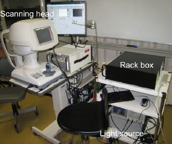
Our colleague Yiheng Lim recently reported a clinical prototype of polarization sensitive optical coherence tomography (PS-OCT) specially designed for the investigation of anterior eye segments. This system based on our developed high-speed Jones matrix OCT methodology, and integrated with a scanning head specially designed for anterior eye imaging. This clinical prototype provides semi-automatic alignment function and possesses a compact, office-compatible size. Several clinical cases were investigated to prove the clinical utility.

The details are reported in Biomedical Optics Express.

>> Full length article on journal web-site (open access)

Citation: Y.H. Lim, M. Yamanari, S. Fukuda, Y. Kaji, T. Kiuchi, M. Miura, T. Oshika, and Y. Yasuno "Birefringence measurement of cornea and anterior segment by office-based polarization-sensitive optical coherence tomography," Biomed. Opt. Express 2, 2392-2402 (2011).

Sunday, April 10, 2011

Journal Article: High-Sensitive Doppler Imaging of Choroid by Dual-Beam Doppler OCT

Our colleague Franck Jaillon recently reported his new Doppler optical coherence tomography (OCT) system which enables high-penetration and dual-velocity imaging of choroidal vasculature. This methodology employed   newly developed dual-beam OCT scanning head to provide high-sensitive Doppler imaging. Furthermore, this scanning head can provide multiple velocity range for Doppler detection. Since this system is using 1-um light as a probe, the details of in vivo choroidal vasculature was successfully visualized.

The details are published in Biomedical Optics Express.

>> Full length article on journal web-site (open access)

Citation: F. Jaillon, S. Makita, E.-J. Min, B.H. Lee, Y. Yasuno, "Enhanced imaging of choroidal vasculature by high-penetration and dual-velocity optical coherence tomography," Biomed. Opt. Express 2, 1147-1158 (2011).

Thursday, October 14, 2010

Journal Article: Wettability Characterization by OCT

Tapio Fabritius (Oulu University, Finland) recently published a paper in Optics Express in which the authors investigated the utility of optical coherence tomography (OCT) for the characterization of wettability of materials. The study presented was a collaboration between Oulu University and Computational Optics Group at the University of Tsukuba.
In the particular demonstration in this paper, a glycerol droplet on a rough surface was characterized, and the contact angles between them were automatically obtained.

The details are published in the following Optics Express paper.

Wednesday, June 16, 2010

Journal Article: Full Range Polarization Sensitive OCT at 1-Micron

Our colleague Masahiro Yamanari recently reported a new polarization sensitive optical coherence tomography (PS-OCT) for retinal and choroidal imaging. This new PS-OCT utilized a probe beam with 1-micron wavelength enabled high-penetration to the choroid, and a full range measurement scheme based on the modulation of reference beam of OCT (BM-scan). This system enables detailed investigation of birefringence property of the deep posterior eye.
After all, this OCT possesses a depth measurement range of 5.3 micrometers in air, a depth resolution of 11.4 micrometers in tissue, and a measurement speed of 30,000 depth-scan/sec. The intensity ratio between an OCT signal and its suppressed mirror signal was found to be 40.5 dB.
This PS-OCT is the first demonstration of 1-um PS-OCT with a clinically useful depth measurement range.
The details are available in the following Optics Express paper.

Citation: M. Yamanari, S. Makita, Y. Lim, and Y. Yasuno, "Full-range polarization-sensitive swept-source optical coherence tomography by simultaneous transversal and spectral modulation," Opt. Express 18, 13964-13980 (2010).

Thursday, April 29, 2010

Adaptive Optics Optical Coherence Tomography at One-Micron

Our colleague Kazuhiro Kurokawa recently reported adaptive optics optical coherence tomography (AO-OCT) for posterior eye imaging. This AO-OCT uses one-micrometer wavelength probe, and is the world first demonstration of high-penetration AO-OCT.
In vivo measurement of human normal subject was performed. The OCT image clearly shows the crosssections of optic nerve fiber bundles as well as the interface between the choroid and sclera.

The details are reported in Optics Express.

Saturday, January 23, 2010

Photonics West Bigins

The biggest annual event of OCT community begins. BiOS, a part of SPIE Photonics West starts on Jun. 23rd. in San Francisco. Almost all members of COG are now rushing into that city. We have 7 oral presentations including 6 from our core member and 1 from our collaborator. See this page to see the abstracts and add our presentations to your conference agenda. Come and see our talks!

Saturday 23rd
  • [2:00pm-2:15pm, Room 306] S. Makita et al., "High-sensitive blood flow imaging of the retina and choroid by using double-beam optical coherence angiography."
  • [5:00pm-5:15pm, Room 306] Y. Lim et al., "Polarization Sensitive Corneal and Anterior Segment Swept-Source Optical Coherence Tomography."
  • [5:15pm-5:30pm, Room 306] A. Miyazawa et al., "Tissue discrimination in anterior eye using three optical parameters obtained by polarization sensitive optical coherence tomography."
Sunday 24th
  • [5:30pm-5:45pm, Room 306] Y. Ikuno et al., "Clinical application of high-penetration optical coherence tomography using 1060-nm wevelength."
Monday 25th
  • [1:00pm-1:15pm, Room 303] K. Kurokawa et al., "Adaptive optics spectral domain optical coherence tomography with one-micrometer light source."
Tuesday 26th
  • [11:15am-11:30am] F. Jaillon et al., "BM-mode scanning with parabolic phase modulation for full range Doppler optical tomography."
Wednesday 27th
  • [8:45am-9:00am] M. Yamanari et al., "Full range polarization-sensitive swept-source optical coherence tomography at 1-um with polarization modulation and BM-mode scan."

Thursday, January 21, 2010

One-Micrometer Adaptive Optics for Scanning Laser Ophthalmoscope


Our colleague Kazuhiro Kurokawa recently reported an adaptive optics scanning laser ophthalmoscope (AO-SLO) with a one-micrometer probe beam. This AO-SLO is the world first demonstration of an adaptive-optics based high-resolution retinal imager with this wavelength band. The mosaic of cone photoreceptors and the flow of blood cells of in vivo human eyes were visualized. The details were published in Optics Express.

Thursday, January 14, 2010

Full-Range Doppler OCT with High Dynamic Range

Franck Jaillon from COG in the University of Tsukuba demonstrated a new full range spectral domain optical coherence tomography (OCT), so called parabolic BM-scan method. This method is a variation of full-range OCT based BM-scan method, but possesses higher tolerance to the sample motion and higher dynamic range of Doppler measurement. In this paper, in vivo investigation of human eye was performed, and it was found that this parabolic BM-scan method is useful for office-based in vivo measurements. The details of this method were published in Optics Express.

Wednesday, January 13, 2010

Yasuno became associate editor of Optics Express

Our colleague Yoshiaki Yasuno became an associate editor of Optics Express. Optics Express is organized by Optical Society of America, and is one of the highest impact journals in the field of optics. Yasuno edits papers mainly of biomedical optics.