Monday, December 26, 2011
Journal paper: Choroidal optical coherence angiography with variable velocity range
The details are described in the following paper.
>> Full length article on Journal web-site (open access)
Citation: F. Jaillon, S. Makita, Y. Yasuno, "Variable velocity range imaging of the choroid with dual-beam optical coherence angiography," Opt. Express 20, 385-396 (2012).
Tuesday, July 26, 2011
Journal Article: Office-Based Polarization Sensitive OCT for Anterior Eye
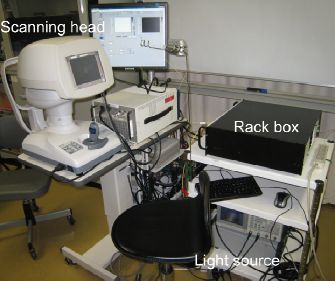
Our colleague Yiheng Lim recently reported a clinical prototype of polarization sensitive optical coherence tomography (PS-OCT) specially designed for the investigation of anterior eye segments. This system based on our developed high-speed Jones matrix OCT methodology, and integrated with a scanning head specially designed for anterior eye imaging. This clinical prototype provides semi-automatic alignment function and possesses a compact, office-compatible size. Several clinical cases were investigated to prove the clinical utility.The details are reported in Biomedical Optics Express.
>> Full length article on journal web-site (open access)
Citation: Y.H. Lim, M. Yamanari, S. Fukuda, Y. Kaji, T. Kiuchi, M. Miura, T. Oshika, and Y. Yasuno "Birefringence measurement of cornea and anterior segment by office-based polarization-sensitive optical coherence tomography," Biomed. Opt. Express 2, 2392-2402 (2011).
Sunday, April 10, 2011
Journal Article: High-Sensitive Doppler Imaging of Choroid by Dual-Beam Doppler OCT
The details are published in Biomedical Optics Express.
>> Full length article on journal web-site (open access)
Citation: F. Jaillon, S. Makita, E.-J. Min, B.H. Lee, Y. Yasuno, "Enhanced imaging of choroidal vasculature by high-penetration and dual-velocity optical coherence tomography," Biomed. Opt. Express 2, 1147-1158 (2011).
Thursday, October 14, 2010
Journal Article: Wettability Characterization by OCT
Tapio Fabritius (Oulu University, Finland) recently published a paper in Optics Express in which the authors investigated the utility of optical coherence tomography (OCT) for the characterization of wettability of materials. The study presented was a collaboration between Oulu University and Computational Optics Group at the University of Tsukuba.Wednesday, June 16, 2010
Journal Article: Full Range Polarization Sensitive OCT at 1-Micron
Our colleague Masahiro Yamanari recently reported a new polarization sensitive optical coherence tomography (PS-OCT) for retinal and choroidal imaging. This new PS-OCT utilized a probe beam with 1-micron wavelength enabled high-penetration to the choroid, and a full range measurement scheme based on the modulation of reference beam of OCT (BM-scan). This system enables detailed investigation of birefringence property of the deep posterior eye.Thursday, April 29, 2010
Adaptive Optics Optical Coherence Tomography at One-Micron
Our colleague Kazuhiro Kurokawa recently reported adaptive optics optical coherence tomography (AO-OCT) for posterior eye imaging. This AO-OCT uses one-micrometer wavelength probe, and is the world first demonstration of high-penetration AO-OCT. Saturday, January 23, 2010
Photonics West Bigins
The biggest annual event of OCT community begins. BiOS, a part of SPIE Photonics West starts on Jun. 23rd. in San Francisco. Almost all members of COG are now rushing into that city. We have 7 oral presentations including 6 from our core member and 1 from our collaborator. See this page to see the abstracts and add our presentations to your conference agenda. Come and see our talks!- [2:00pm-2:15pm, Room 306] S. Makita et al., "High-sensitive blood flow imaging of the retina and choroid by using double-beam optical coherence angiography."
- [5:00pm-5:15pm, Room 306] Y. Lim et al., "Polarization Sensitive Corneal and Anterior Segment Swept-Source Optical Coherence Tomography."
- [5:15pm-5:30pm, Room 306] A. Miyazawa et al., "Tissue discrimination in anterior eye using three optical parameters obtained by polarization sensitive optical coherence tomography."
- [5:30pm-5:45pm, Room 306] Y. Ikuno et al., "Clinical application of high-penetration optical coherence tomography using 1060-nm wevelength."
- [1:00pm-1:15pm, Room 303] K. Kurokawa et al., "Adaptive optics spectral domain optical coherence tomography with one-micrometer light source."
- [11:15am-11:30am] F. Jaillon et al., "BM-mode scanning with parabolic phase modulation for full range Doppler optical tomography."
- [8:45am-9:00am] M. Yamanari et al., "Full range polarization-sensitive swept-source optical coherence tomography at 1-um with polarization modulation and BM-mode scan."
Thursday, January 21, 2010
One-Micrometer Adaptive Optics for Scanning Laser Ophthalmoscope

Our colleague Kazuhiro Kurokawa recently reported an adaptive optics scanning laser ophthalmoscope (AO-SLO) with a one-micrometer probe beam. This AO-SLO is the world first demonstration of an adaptive-optics based high-resolution retinal imager with this wavelength band. The mosaic of cone photoreceptors and the flow of blood cells of in vivo human eyes were visualized. The details were published in Optics Express.
Thursday, January 14, 2010
Full-Range Doppler OCT with High Dynamic Range
Franck Jaillon from COG in the University of Tsukuba demonstrated a new full range spectral domain optical coherence tomography (OCT), so called parabolic BM-scan method. This method is a variation of full-range OCT based BM-scan method, but possesses higher tolerance to the sample motion and higher dynamic range of Doppler measurement. In this paper, in vivo investigation of human eye was performed, and it was found that this parabolic BM-scan method is useful for office-based in vivo measurements. The details of this method were published in Optics Express.Wednesday, January 13, 2010
Yasuno became associate editor of Optics Express
Friday, January 8, 2010
Generalized Jones Matrix OCT
Shuichi Makita and his colleagues in COG recently reported the generalization of Jones matrix optical coherence tomography (Jones OCT) in Optics Express. In this paper, we generalized theoretical model of Jones OCT which provides us not only phase retardation tomography but local birefringence tomography of biological and medical samples. The noise property of this generalized Jones OCT is also carefully analyzed. This method was applied to visualize the details of an anterior eye chamber, and successfully contrast a trabecular meshwork. The details are described in the following paper.Thursday, January 7, 2010
Investigation of Choroidal Thickness

Our collaborator Yasushi Ikuno (Osaka University Hospital) investigated the choroidal thickness of healthy Japanese. The chorid is one of the tissues in the eye ant it locates beneath the retina, and which had not been possible to be clearly visualized by OCT. He investigated the choroid by using a brand new high-penetration OCT made by our group (COG, Tsukuba). After the examination of 86 eyes, he found that the choroidal thickness varies around its position, and is associated the refractive error of the eye, axial eye length, and the age of the subject. The details are reported in Investigative Ophthalmology and Visual Science (IOVS, a journal of association for research in vision and ophthalmology).
doi:10.1167/iovs.09-4383
Birefringence Imaging of Necrotizing Scleritis

Our colleagues Masahiro Miura published a case report of necrotizing scleritis, which is an inflammation disease of sclera. The anterior eye of a patient was scanned by our anterior eye segment polarization sensitive optical coherence tomography prototype (AS-PS-OCT). The tissue selective imaging ability of AS-PS-OCT visualized the abnormal thinning of the eye. The details were published in Ophthalmic Surgery, Lasers and Imaging.
Wednesday, September 16, 2009
Tissue Discrimination Imaging of Anterior Eye

Our colleague Arata Miyazawa reported a new imaging methodology which used polarization sensitive optical coherence tomography (PS-OCT) and a newly developed algorithm to discriminate tissues based on their optical properties. This algorithm uses three optical properties obtained by PS-OCT including scattering intensity, signal extinction coefficient along depth, and local birefringence, and discriminates tissues based on these properties. By using this algorithm, five tissue types of an anterior eye including trabecular meshwork, conjunctiva, sclera, cornea and uvea were successfully discriminated. The details was published in Optics Express.
Friday, September 11, 2009
Ocular Biometry by 3D-OCT

Our collaborator Shinichi Fukuda investigated the utility of 3D corneal and anterior eye segment optical coherence tomography (3D-CAS-OCT) for ocular biometry. The ocular biometry is a method to extract numerical parameters from an eye, which represent the functions and integrity of the eye, e.g. a central corneal thickness (CCT) or an anterior eye chamber depth (ACD).
He examined 40 eyes of 40 normal subjects by 3D-CAS-OCT, Scheimpflug camera, scanning-slit topography and ultrasonic pachymetry, and obtained CCT, ACD and other parameters. This study indicated 3D-CAS-OCT was usable for ocular biometry.
This study has been reported on a journal Ophthalmology. The paper and abstract is available on the following locations.
>> Journal web site
>> Request reprint via e-mail (please indicate the following citation in the e-mail.)
Citation: S. Fukuda, K. Kawana, Y. Yasuno and T. Oshika, "Anterior ocular biometry using 3-dimensional optical coherence tomography," Ophthalmology 116, 882-889 (2009).